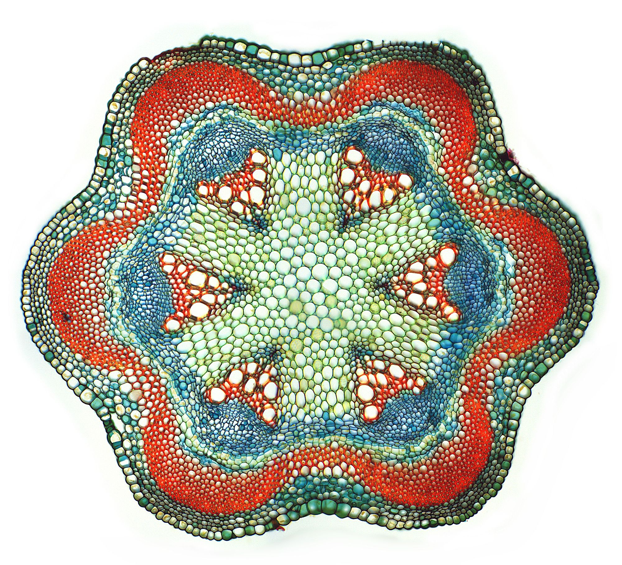
Your Cross section of an animal cell images are available in this site. Cross section of an animal cell are a topic that is being searched for and liked by netizens today. You can Get the Cross section of an animal cell files here. Get all royalty-free images.
If you’re looking for cross section of an animal cell images information connected with to the cross section of an animal cell interest, you have visit the right site. Our website always provides you with hints for seeing the highest quality video and image content, please kindly surf and locate more informative video content and images that fit your interests.
Cross Section Of An Animal Cell. • you will see a cell like this on your quiz next week. Includes model and activity guide. Animal cells lack cell walls. The largest known animal cell is the ostrich egg, which can stretch over 5.1 inches across and weighs about 1.4 kilograms.
 Animal Cell model (With images) Cells project, Animal From pinterest.com
Examine the animal cell diagram and recognize parts like the centrioles, lysosomes, golgi bodies, ribosomes and more indicated clearly. • you will see a cell like this on your quiz next week. Cross section cut under the microscope microscopic view of animal cells as seen in the fluorescence microscope after fixation dyi microscope image. • remember there are no “flying” labels. • remember there are no “flying” labels. They should be parallel to the bottom of the page.
This is an online quiz called cross section of an animal cell there is a printable worksheet available for download here so you can take the quiz with pen and paper.
It controls many of the functions of the. What scale factor was used to draw the diagram? Find the perfect cell cross section stock illustrations from getty images. Cut & glue into your notebook vacuole. The other features only letters next to each cell part, making it a natural tool for your child to practice for his next test at home. More stock illustrations from this artist see all.
 Source: pinterest.com
Cell structure, cross section on an abstract blue science background with transparent dna chains anatomy of the cell cross section beautiful colorful illustration on a light blue abstract background So, i�m always on the lookout for inexpensive models of things that will be studied again and again, and was very excited to find these! In fact, the cell�s diameter is 0.15 mm. Animal cell, bacterial cell and plant cell structure, cross section detailed colorful anatomy on bright gradient plant cellulose biology vector illustration diagram. • remember there are no “flying” labels.
 Source: in.pinterest.com
Plant cell parts animal cell organelles plant cell diagram animal cell project plant and animal cells shapes worksheet kindergarten map activities educational activities cell model more information. Examine the animal cell diagram and recognize parts like the centrioles, lysosomes, golgi bodies, ribosomes and more indicated clearly. Animal cells lack cell walls. Animal cell and plant cell line In fact, the cell�s diameter is 0.15 mm.
 Source: pinterest.com
This is an online quiz called cross section of an animal cell there is a printable worksheet available for download here so you can take the quiz with pen and paper. Animal cell and plant cell line Tissues, organs, and organ systems enabled the evolution of large, multicellular bodies. Color each organelle a different color. A copy is made using a scale factor of 150%.
 Source: pinterest.com
Tissues are necessary to produce organs and organ systems. Golgi apparatus a part of the eukaryotic cell. The soft foam cell splits in half to show the key parts of an animal cell, including the nucleus, nucleolus, vacuole, centrioles, cell membrane and more. • remember there are no “flying” labels. They should be parallel to the bottom of the page.
 Source: pinterest.com
What scale factor was used to draw the diagram? Tissues, organs, and organ systems enabled the evolution of large, multicellular bodies. Cut & glue into your notebook vacuole. Plant cell parts animal cell organelles plant cell diagram animal cell project plant and animal cells shapes worksheet kindergarten map activities educational activities cell model more information. One hemisphere is labeled with the parts of the cell;
 Source: pinterest.com
• remember there are no “flying” labels. Golgi apparatus a part of the eukaryotic cell. Except for sponges, animal cells are arranged into tissues. Includes model and activity guide. Cut & glue into your notebook vacuole.
 Source: pinterest.com
A copy is made using a scale factor of 150%. The largest known animal cell is the ostrich egg, which can stretch over 5.1 inches across and weighs about 1.4 kilograms. (2 marks) b) what is the A copy is made using a scale factor of 150%. This is an online quiz called cross section of an animal cell there is a printable worksheet available for download here so you can take the quiz with pen and paper.
 Source: pinterest.com
A) what are the dimensions of the copy? • remember there are no “flying” labels. They should be parallel to the bottom of the page. It controls many of the functions of the. A) what are the dimensions of the copy?
 Source: pinterest.com
Examine the animal cell diagram and recognize parts like the centrioles, lysosomes, golgi bodies, ribosomes and more indicated clearly. • you will see a cell like this on your quiz next week. Select from premium cell cross section images of the highest quality. The other features only letters next to each cell part, making it a natural tool for your child to practice for his next test at home. A snapshot of supplies used to make a cross section of an animal cell a volunteer and i created our model in a few hours.
 Source: pinterest.com
A photograph is 6 cm by 11 cm. • remember there are no “flying” labels. This is where the digestion of cell nutrients takes place. Animal cell and plant cell structure, cross section detailed colorful anatomy. • remember there are no “flying” labels.
 Source: pinterest.com
Anatomy of the cell cross section beautiful colorful illustration on a light blue abstract background; They should be parallel to the bottom of the page. The soft foam cell splits in half to show the key parts of an animal cell, including the nucleus, nucleolus, vacuole, centrioles, cell membrane and more. Except for sponges, animal cells are arranged into tissues. If you make one, please know that the longest part is gathering and arranging the supplies.
 Source: pinterest.com
Animal cell cross section structure of a eukaryotic cell vector diagram; Cut & glue into your notebook vacuole. The soft foam cell splits in half to show the key parts of an animal cell, including the nucleus, nucleolus, vacuole, centrioles, cell membrane and more. This is an online quiz called cross section of an animal cell there is a printable worksheet available for download here so you can take the quiz with pen and paper. Examine the animal cell diagram and recognize parts like the centrioles, lysosomes, golgi bodies, ribosomes and more indicated clearly.
 Source: pinterest.com
Cut & glue into your notebook vacuole. Animal cells lack cell walls. Tissues are necessary to produce organs and organ systems. Color each organelle a different color. Cut & glue into your notebook vacuole.
 Source: pinterest.com
Cell structure, cross section on an abstract blue science background with transparent dna chains anatomy of the cell cross section beautiful colorful illustration on a light blue abstract background It controls many of the functions of the. A copy is made using a scale factor of 150%. Animal cell and plant cell structure, cross section detailed colorful anatomy. One hemisphere is labeled with the parts of the cell;
 Source: pinterest.com
Find the perfect cell cross section stock illustrations from getty images. The other features only letters next to each cell part, making it a natural tool for your child to practice for his next test at home. What scale factor was used to draw the diagram? This is in stark contrast to the neuron in the human body, which is just 100 microns across. Animal cell cross section structure of a eukaryotic cell vector diagram;
 Source: pinterest.com
A photograph is 6 cm by 11 cm. What scale factor was used to draw the diagram? Animal cell cross section structure of a eukaryotic cell vector diagram; Except for sponges, animal cells are arranged into tissues. A snapshot of supplies used to make a cross section of an animal cell a volunteer and i created our model in a few hours.
 Source: pinterest.com
• remember there are no “flying” labels. Animal cell and plant cell structure, cross section detailed colorful anatomy. Animal cell, bacterial cell and plant cell structure, cross section detailed colorful anatomy on bright gradient plant cellulose biology vector illustration diagram. This is in stark contrast to the neuron in the human body, which is just 100 microns across. The other features only letters next to each cell part, making it a natural tool for your child to practice for his next test at home.
 Source: pinterest.com
A photograph is 6 cm by 11 cm. In the diagram, the diameter of the cell is 4.5 cm. Examine the animal cell diagram and recognize parts like the centrioles, lysosomes, golgi bodies, ribosomes and more indicated clearly. A copy is made using a scale factor of 150%. Animal cell cross section structure of a eukaryotic cell vector diagram;
This site is an open community for users to submit their favorite wallpapers on the internet, all images or pictures in this website are for personal wallpaper use only, it is stricly prohibited to use this wallpaper for commercial purposes, if you are the author and find this image is shared without your permission, please kindly raise a DMCA report to Us.
If you find this site helpful, please support us by sharing this posts to your preference social media accounts like Facebook, Instagram and so on or you can also save this blog page with the title cross section of an animal cell by using Ctrl + D for devices a laptop with a Windows operating system or Command + D for laptops with an Apple operating system. If you use a smartphone, you can also use the drawer menu of the browser you are using. Whether it’s a Windows, Mac, iOS or Android operating system, you will still be able to bookmark this website.


